By Georgia Barrington-Smith & Dr Rebecca Duncan
For decades, conventional X-rays have been invaluable in clinical settings, enabling doctors and radiographers to gain critical insights into patients’ health. While traditional X-rays are still widely used, they are limited in the depth of information they can provide. New, advanced multimodal techniques, like phase-shift and dark-field imaging, can offer more detailed images, revealing not only bones and metal implants but also microstructures and surrounding soft tissue.
Unlike conventional X-ray imaging, which focuses on the absorption of X-rays by the sample (attenuation), phase-shift imaging captures changes in the phase of X-rays as they pass through the sample. In addition, dark-field imaging highlights small structures such as tiny pores, cracks, or granular textures, providing detailed information beyond the spatial resolution of traditional X-rays.
Enhancing X-ray imaging efficiency with multimodal techniques
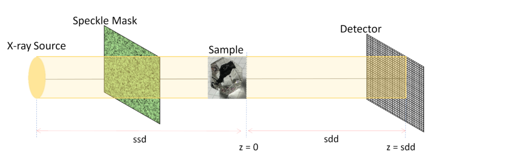
Research into multimodal techniques has introduced one promising method: Speckle-Based X-ray Imaging (SBXI). With a simple experimental setup and ability to produce high-quality images with minimal data, SBXI uses a spatially-varied medium (such as sandpaper or textured materials) placed between the X-ray source and detector. This creates speckles—tiny variations in the intensity of the X-rays as they pass through the sample. These speckles act as markers, helping to track the X-ray wavefronts. By analysing how the speckles change as they pass through the sample, additional structural information can be recovered.
Although SBXI shows great potential, there are significant challenges hindering its development, such as the longer time required to construct high-quality images. Therefore, fast, efficient computer-based algorithms are crucial not only for reducing computational time, but also for minimising the radiation exposure to the patient during image reconstruction.
Samantha’s steps towards speckle-based X-rays
Samantha Alloo, an AINSE PGRA scholar, and her collaborators at ANSTO and the University of Canterbury have developed a fast, computationally efficient algorithm capable of reconstructing multimodal signals in just a few seconds. This new algorithm, called Multimodal Intrinsic Speckle-Tracking (MIST), provides on-demand multimodal imaging with low radiation exposure to the patient, or other delicate samples.
MIST uses the principle of energy conservation at small scales to track speckles and generate detailed images in Speckle-Based X-ray Imaging. The dark-field images it produces are especially useful when combined with other traditional X-ray imaging methods, such as Small-Angle X-ray Scattering (SAXS), as they can reveal information about sample structures beyond the limits of the imaging system’s spatial resolution. This capability has already proven valuable in applications including clinical mammography, biosecurity, and engineering crack detection.
Future directions for Samantha’s algorithm
Samantha and her team aim to further develop MIST to create a user-friendly SBXI setup at the MicroCT beamline at ANSTO’s Australian Synchrotron. This setup is designed to retrieve high-quality data comparable to, or even surpassing, well-established imaging techniques. The goal is to make these algorithms computationally efficient and general enough to be applied in a wide range of real-time synchrotron and laboratory experiments.
Samantha hopes to extend the MIST algorithm to work with materials whose properties vary depending on the direction that X-rays travel through the material. This will allow the reconstructed dark-field signal to reveal information about the sample’s microstructure orientation, making MIST suitable for a broader range of samples. This expanded approach will contribute to the overall goal of creating a more robust and user-friendly technique for clinical applications.
Finally, while utilising the MIST algorithm to conduct SBXI experiments, Samantha also hopes to develop a dark-field tensor tomography protocol. This protocol promises to deliver more detailed information about a sample’s internal structure by using SBXI techniques to capture its orientation in three-dimensional space.
AINSE are proud to spotlight Samantha Alloo for her groundbreaking work!
To explore more incredible research by our AINSE scholars, visit ainse.edu.au/research-spotlight.
We’re hanging up our stethoscopes and turning our attention to Bushfire Awareness in April, where we’ll spotlight two AINSE scholars doing crucial research on Australia’s bushfires and explore how science is driving better outcomes for our community.
Stay up to date with AINSE by following us on all our social media platforms @ainse_ltd on Instagram, Facebook, Threads and LinkedIn.